
Antioxidants and Age-Related Macular Degeneration
It is estimated that 150,000 Americans are legally blind from AMD, with 20,000 new cases occurring per year. In addition to the effects of ultra-violet light, other risk factors for Age-Related Macular Degeneration (AMD) include aging, atherosclerosis, high blood pressure and smoking. (1) Although all the underlying causes of AMD are not fully understood, it has been shown that free radical damage from singlet oxygen caused by ultraviolet light reaching the macula is a significant contributing factor. Added to this are the free radicals generated by the mitochondria, the energy-producing organelles found in large quantities in all the cells of the eye. The eye, like the brain has a high metabolic rate and high bioelectric activity with relatively low antioxidant capacity to deal with reactive oxygen and nitrogen species. The eye is also under carbonyl stress caused by protein oxidation.
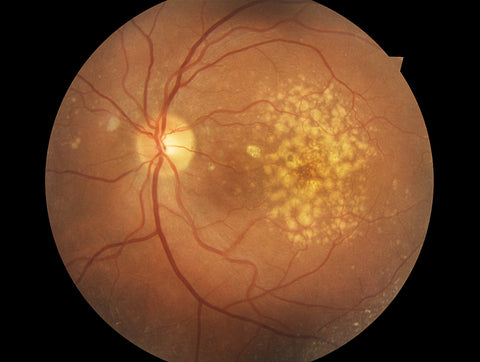 Early AMD is characterized clinically by yellow deposits known as drusen and changes in pigmentation of the retina. Late AMD develops when there is an in-growth of new blood vessels that bleed into the subretinal space (exudative or “wet” type) or when the macula atrophies (geographic atrophy or “dry” type). Both these conditions usually lead to severe loss of central vision.
This entire scenario creates a condition of oxidative stress, defined by Helmut Seis in 1986 as an imbalance between excess free radical generation overwhelming the body’s antioxidant ability to quench free radicals. (2)
The debate about the ability of antioxidants to forestall or prevent the progression of cataracts and AMD has been largely resolved by the AREDS 1 and AREDS 2 studies sponsored by the N.I.H. (3, 4) However, scientific studies published before, between and after these studies demonstrates a wide variety of antioxidants and herbal extracts containing a broad mixture of plant-derived antioxidants indeed does prevent or delay the progression of these eye diseases.
Antioxidants are thought to prevent AMD by reducing the photo-oxidative damage from blue light in the oxygen filled environment of the retina, which is rich in polyunsaturated fatty acids that are highly susceptible to oxidation. (5)
Carotenoids have been shown to be good filters of harmful blue light, and their antioxidative properties have been demonstrated in vitro. (5, 6) Two of these carotenoids, lutein and zeaxanthin, are found in the macula in concentrations higher than in other parts of the body.
The earliest study of antioxidants and AMD took place in France and Germany and used Ginkgo biloba extract which has potent antioxidant properties. It is believed the extract may help to slow down the progression of AMD. (7, 8) Two published trials were identified that randomized a total of 119 people. In one study conducted in France, 20 people were randomly allocated to Gingko biloba extract 80 mg twice daily or placebo. In the other study conducted in Germany, 99 people were randomly allocated to two different doses of Ginkgo biloba extract (240 mg per day and 60 mg per day). Treatment duration in both studies was six months. Both trials reported some positive effects of Ginkgo biloba on vision.
Bilberry extract
Bilberry contains 15 different anthocyanins. Bilberry extract inhibited the generation of intracellular free radical induced by blue LED light irradiation on retinyl photoreceptor cells.
Bilberry has also been reported to improve visual function in animal models and in clinical trials. (10, 11) Animal studies have demonstrated Bilberry extract to be beneficial in preventing retinal inflammation and cataracts. (12, 13)
Vitamin C
The EDCC (Eye Disease Case Control) Study reported that low plasma levels of vitamin C were associated with increased risk of AMD. Furthermore, an antioxidant index, comprising plasma carotenoids, selenium, ascorbate, and vitamin E lowered AMD risk. (14) The Baltimore Longitudinal Study of Aging (BLSA) observed a significant protective effect for an antioxidant index which included a-tocopherol, ascorbate, and beta carotene. (15)
Vitamin E
The retina contains high quantities of vitamin E in the rod outer segments and in the RPE. (16, 17) The concentrations within these tissues are very sensitive to the dietary intake of the vitamin. (18, 19) The retinyl pigment epithelium (RPE) content of vitamin E, which was 4 to 7 times that of the neural retina, increased with increasing age, and that this rise was in response to increasing oxidative stress. (20) It has also been noted that the central neural retina closely regulates its vitamin E content (21) and there are remarkable similarities with the carotenoids of the macular pigment. (22, 23, 24) Interestingly, carotenoids and a-tocopherol can act synergistically as free radical scavengers. (25)
Antioxidant synergy was also seen in the Rotterdam Eye Study, where an above average dietary intake of combined antioxidants similar to those found in the clinical trial formulation of the AREDS study was associated with a larger reduced risk of AMD than for each antioxidant, vitamin C, vitamin E, β-carotene and zinc studied individually. (26)
Shop VisiVite provides premium antioxidant supplements for the support of the retina and macula.

References:
1.Murray, M. and Pizzorno, J. Encyclopedia of Natural Medicine (2nd edit) Prima Health 1998: 319-324.
2. Sies, H. Biochemistry of Oxidative Stress. Vol. 25; 12, Dec 1986, pp. 1058-1071.
3. Age-Related Eye Disease Study Research Group. A randomized, placebo-controlled, clinical trial of high-dose supplementation with vitamins C and E, beta carotene, and zinc for age-related macular degeneration and vision loss: AREDS report No 8. Arch Ophthalmol 2001; 119:1417-36.
4. AREDS2 Research Group, Chew, EY, Clemons, T, et al. The Age-Related Eye Disease Study 2 (AREDS2): study design and baseline characteristics (AREDS2 report number 1). Opthalmology. 2012 Nov;119(11):2282-9.
5.Kirschfeld K. Carotenoid pigments: their possible role in protecting against photooxidation in eyes and photoreceptor cells. Proc R Soc Lond B Biol Sci 1982; 216:71-85.
6. Britton G. Structure and properties of carotenoids in relation to function. Faseb J.1995; 9:1551-8.
7. Evans JR. Ginkgo biloba extract for age-related macular degeneration (Review) Cochrane Database of Systematic Reviews
2013, Issue 1. Art. No.: CD001775.
8. Fies P, Dienel A. [Ginkgo extract in impaired vision – treatment with special extract Egb 761 of impaired vision due to dry senile macular degeneration]. Wiedn Med Wochenschr. 2002;152(15-16):423-6.
9. Ogawa, K, Kuse, Y., et al. Protective effects of bilberry and lingonberry extracts against blue light-emitting diode light-induced retinal photoreceptor cell damage in vitro.BMC Compolement Altern Med. 2014; 14: 120.
10. Miyake S, Takahashi N, Sasaki M, Kobayashi S, Tsubota K, Ozawa Y. Vision preservation during retinal inflammation by anthocyanin-rich bilberry extract: cellular and molecular mechanism. Lab Investig. 2012; 92 :102–109.
11. Matsunaga N, Imai S, Inokuchi Y, Shimazawa M, Yokota S, Araki Y, Hara H. Bilberry and its main constituents have neuroprotective effects against retinal neuronal damage in vitro and in vivo. Mol Nutr Food Res. 2009; 53: 869–877.
12. Matsunaga N, Chikaraishi Y, Shimazawa M, Yokota S, Hara H. Vaccinium myrtillus (bilberry) extracts reduce angiogenesis in vitro and in vivo. Evid Based Complement Alternat Med. 2010; 7: 47–56.
13. Ogawa K, Tsuruma K, Tanaka J, Kakino M, Kobayashi S, Shimazawa M, Hara H. The protective effects of bilberry and lingonberry extracts against UV light-induced retinal photoreceptor cell damage in vitro. J Agric Food Chem. 2013; 61:8.
14. Antioxidant status and neovascular age-related macular degeneration. The Eye Disease Case-Control Study Group. Arch Ophthalmol 111:104–9, 1993.
15. West S, Vitale S, Hallfrisch J, et al: Are antioxidants or supplements protective for age-related macular degeneration? [see comments]. Arch Ophthalmol 112:222–7, 1994.
16. Delcourt C, Cristol JP, Tessier F, et al: Age-related macular degeneration and antioxidant status in the POLA study. POLA Study Group. Pathologies Oculaires Liees a l’Age. Arch Ophthalmol 117:1384–90, 1999.
17. Friedrichson T, Kalbach HL, Buck P, van Kuijk FJ: Vitamin E in macular and peripheral tissues of the human eye. Curr Eye Res 14:693–701, 1995.
18. Hunt DF, Organisciak DT, Wang HM, et al: alpha-Tocopherol in the developing rat retina: a high-pressure liquid chromatographic analysis. Curr Eye Res 3:1281–8, 1984.
19. Stephens RJ, Negi DS, Short SM, et al: Vitamin E distribution in ocular tissues following long-term dietary depletion and supplementation as determined by microdissection and gas chromatography-mass spectrometry. Exp Eye Res 47:237–45, 1988.
20. Organisciak DT, Berman ER, Wang HM, Feeney-Burns L: Vitamin E in human neural retina and retinal pigment epithelium: effect of age. Curr Eye Res 6:1051–5, 1987.
21. Crabtree DV, Adler AJ, Snodderly DM: Vitamin E, retinyl palmitate, and protein in rhesus monkey retina and retinal pigment epithelium-choroid. Invest Ophthalmol Vis Sci 37: 47–60, 1996.
22. Handelman GJ, Snodderly DM, Krinsky NI, et al: Biological control of primate macular pigment. Biochemical and densitometric studies. Invest Ophthalmol Vis Sci 32:257–67, 1991.
23. Snodderly DM, Auran JD, Delori FC: The macular pigment. II. Spatial distribution in primate retinas. Invest Ophthalmol Vis Sci 25:674–85, 1984.
24. Snodderly DM, Handelman GJ, Adler AJ: Distribution of individual macular pigment carotenoids in central retina of macaque and squirrel monkeys. Invest Ophthalmol Vis Sci 32:268–79, 1991.
25. Palozza P, Krinsky NI: beta-Carotene and alpha-tocopherol are synergistic antioxidants. Arch Biochem Biophys 297: 184–7, 1992.
26.Ikram, MA, Bruselle, GG, et al. The Rotterdam Study: 2018 update on objectives, design and main results. Eur J Epidemiol. 2017; 32(9): 807–850.